What is trachea midline?
Keeping this in view, what is the meaning of trachea?
Listen to pronunciation. (TRAY-kee-uh) The airway that leads from the larynx (voice box) to the bronchi (large airways that lead to the lungs). Also called windpipe.
Similarly, what does it mean if the trachea is not midline? Tracheal deviation is most commonly caused by injuries or conditions that cause pressure to build up in your chest cavity or neck. Openings or punctures in the chest wall, the lungs, or other parts of your pleural cavity can cause air to only move in one direction inward.
Beside this, what is in front of the trachea?
In front of the upper trachea lies connective tissue and skin. Several other structures pass over or sit on the trachea; the jugular arch, which joins the two anterior jugular veins, sits in front of the upper part of the trachea. The sternohyoid and sternothyroid muscles stretch along its length.
Which of these is the best definition for trachea?
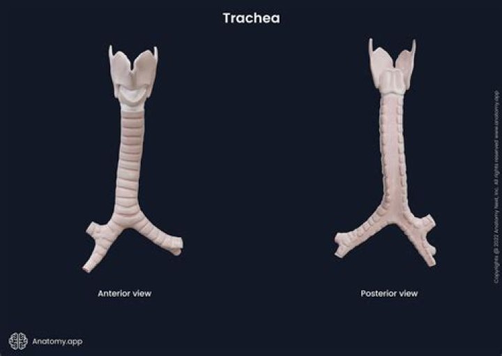
The trachea, commonly known as the windpipe, is a tube about 4 inches long and less than an inch in diameter in most people. The trachea begins just under the larynx (voice box) and runs down behind the breastbone (sternum). The trachea then divides into two smaller tubes called bronchi: one bronchus for each lung.
Related Question Answers
What is trachea and its function?
The trachea serves as passage for air, moistens and warms it while it passes into the lungs, and protects the respiratory surface from an accumulation of foreign particles. The trachea is lined with a moist mucous-membrane layer composed of cells containing small hairlike projections called cilia.What happens to the windpipe or trachea before it reaches the lungs?
When you breathe in (inhale), air containing oxygen enters your windpipe, passes through the bronchi and eventually reaches the air sacs. These air sacs, called alveoli, are responsible for gas exchange. They look a bit like grapes at the end of the bronchial branches.What connects the lungs to the trachea?
At its bottom end, the trachea divides into left and right air tubes called bronchi (BRAHN-kye), which connect to the lungs.What is the root word for trachea?
The word trachea is from the Greek phrase for windpipe — trakheia arteria, which literally meant "rough artery." The trachea is formed from rings of cartilage, which give the trachea its rough appearance.What is other name of windpipe?
Also called trachea. Enlarge. Anatomy of the respiratory system, showing the trachea and both lungs and their lobes and airways.What is another name for larynx?
voice boxWhich structure is more anterior the esophagus or the trachea?
The trachea descends anterior to the esophagus, enters the superior mediastinum, and divides into right and left main bronchi. The trachea is a median structure but, near its lower end, deviates slightly to the right, resulting in the left main bronchus crossing anterior to the esophagus.Can you live without a trachea?
Thomas was born without a trachea — the cartilaginous tube through which we breathe. The condition is called tracheal agenesis, and it is extremely rare. Fewer than 200 cases have been identified in more than a century. The lifespan of an infant born without a trachea is measured in minutes.Why is the trachea deviated to the right?
The trachea is generally a midline structure displaced slightly to the right by the aortic arch. Various conditions, including mediastinal masses and vascular anomalies, may bow, displace or indent the trachea. Such appearances are most commonly seen in patients with thyroid masses or a right-sided aortic arch.What would happen if the trachea didn't have cartilaginous rings?
TO MAINTAIN THE TRACHEA IN ITS WIDE OPEN STATE DURING INHALATION. IF WE DIDN'T HAVE THE C-SHAPED CARTILAGINOUS RINGS HOLDING THE TRACHEA OPEN, DURING INHALATION THE TRACHEA WOULD COLLAPSE.What happens if the trachea is damaged?
Windpipe injuriesSometimes the swelling can be extensive enough that it can actually start to block off the airway,” Stankus said. “If you have any rapid breathing or difficulty breathing, changes to your voice, wheezing (stridor), or odd changes in the sound of your breathing,” it's an emergency, Stankus said.
Why does my trachea hurt?
Causes. Bacterial tracheitis is a bacterial infection of the trachea and is capable of producing airway obstruction. One of the most common causes is Staphylococcus aureus and often follows a recent viral upper respiratory infection. Bacterial tracheitis is a rare complication of influenza infection.Is the esophagus in front of the trachea?
The esophagus is a muscular tube connecting the throat (pharynx) with the stomach. The esophagus is about 8 inches long, and is lined by moist pink tissue called mucosa. The esophagus runs behind the windpipe (trachea) and heart, and in front of the spine.Why does the trachea have cilia?
The walls of the trachea (pronounced: TRAY-kee-uh) are strengthened by stiff rings of cartilage to keep it open. The trachea is also lined with cilia, which sweep fluids and foreign particles out of the airway so that they stay out of the lungs.When we inhale we breathe in air into the lungs What do we breathe out when we exhale?
We get oxygen by breathing in fresh air, and we remove carbon dioxide from the body by breathing out stale air. But how does the breathing mechanism work? Air flows in via our mouth or nose. The air then follows the windpipe, which splits first into two bronchi: one for each lung.Why is the trachea C shaped?
The cartilaginous rings are C-shaped to allow the trachea to collapse slightly at the opening so that food can pass down the esophagus. The esophagus lies posteriorly to the trachea. The mucocilliary escalator helps prevent pathogens from entering the lungs.Is your trachea supposed to move?
The normal position of the trachea is straight up and down, running along the center of the front side of the throat. Certain conditions can cause the trachea to shift to one side or the other.What is the difference between Oesophagus and trachea?
The esophagus is the tube that connects the throat to the stomach. The trachea is the tube that connects the throat to the windpipe and lungs. Normally, the esophagus and trachea are two tubes that are not connected.Can the heart shift?
Sometimes, your heart develops pointing the wrong way because other anatomical problems exist. Defects in your lungs, abdomen, or chest can cause your heart to develop so that it's shifted towards the right side of your body.What could happen if air gets caught in between the space between the lungs and thoracic cavity?
A collapsed lung, also known as a pneumothorax, is a condition that occurs when air enters the space between the chest wall and the lung (pleural space). As air builds up, pressure inside the pleural space increases and causes the lung to collapse.Does a chest xray show the trachea?
On chest radiographs, the distal cervical trachea, intrathoracic trachea and main bronchi are visible; however, overlying mediastinal structures often obscure intrathoracic tracheobronchial abnormalities.Which way does the trachea deviate in tension pneumothorax?
Tension pneumothorax is classically characterized by hypotension and hypoxia. On examination, breath sounds are absent on the affected hemothorax and the trachea deviates away from the affected side. The thorax may also be hyperresonant; jugular venous distention and tachycardia may be present.Who is at risk for spontaneous pneumothorax?
In most cases of spontaneous pneumothorax, the cause is unknown. Tall and thin adolescent males are typically at greatest risk, but females can also have this condition. Other risk factors include connective tissue disorders, smoking, and activities such as scuba diving, high altitudes and flying.How does bacterial pneumonia lead to hypoxemia?
Arterial hypoxemia early in acute pneumococcal pneumonia is principally caused by persistence of pulmonary artery blood flow to consolidated lung resulting in an intrapulmonary shunt, but also, to a varying degree, it is caused by intrapulmonary oxygen consumption by the lung during the acute phase and by ventilation-What is the difference between a pneumothorax and a tension pneumothorax?
By allowing the air to escape, the pneumothorax does not get any larger and the pressure can't build and transform the injury into a tension pneumothorax. Tension pneumothorax is a life-threating process that needs emergent treatment.When palpating trachea position it should be?
The trachea should be checked to see if it is in the normal central position. This means the distance between the trachea and the sternomastoid muscles should be equal on both sides. Slight displacement of the trachea to the right is fairly common in healthy people (Talley and O'Connor, 2001).How do you palpate the trachea?
Ask the patient to relax the sternomastoid muscles by dropping their chin, and to lean slightly forward. Rest your middle finger on the suprasternal notch and pass it on either side of the trachea as deeply and inferiorly as possibly (figure 14a,b).How do you assess the position of the trachea?
Method Of ExamInspect for the symmetry of clavicular insertion of both sternomastoids. Tracheal Position: Gently bend the head to relax the sternomastoids. By inserting your finger between the trachea and sternomastoid, assess and compare the space on either side.